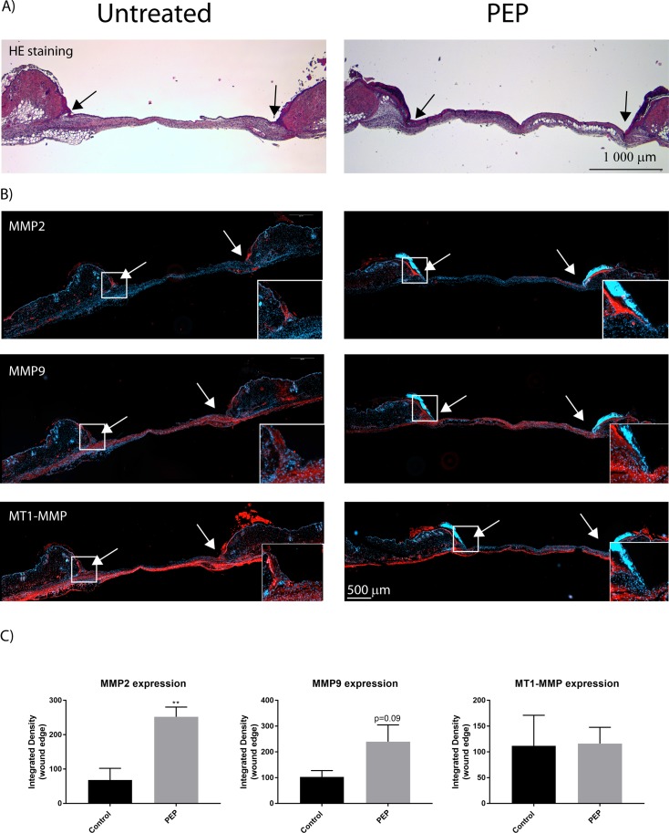
Fig 9. MMP-2 and MMP-9, but not MT1-MMP protein expression are elevated at wound edges with PEP treatment by day 3.
A) H&E staining showing the contour of excisional wound sections at day 3. B) Immunofluorescence of excisional wound sections at day 3 with antibodies targeting MMP-2, MMP-9, and MT1-MMP (red). The cell nuclei are counterstained with Hoechst (blue). Arrows show the wound edges. Inserts show magnification of boxed areas for the wound edges. C) Integrated densities of selected areas at wound edges at day 3, as presented in inserts in B were quantified using ImageJ. Presented values are Integrated Densities (sum of pixel values in selection * area of selection). Both sides of the wound edge were quantified for each treatment (n = 3 mice). Asterisk indicate significant differences (**p<0.01 in PEP treated mice compared with control mice) statistics assessed by unpaired two-tailed t-test.

