Fig. 3.
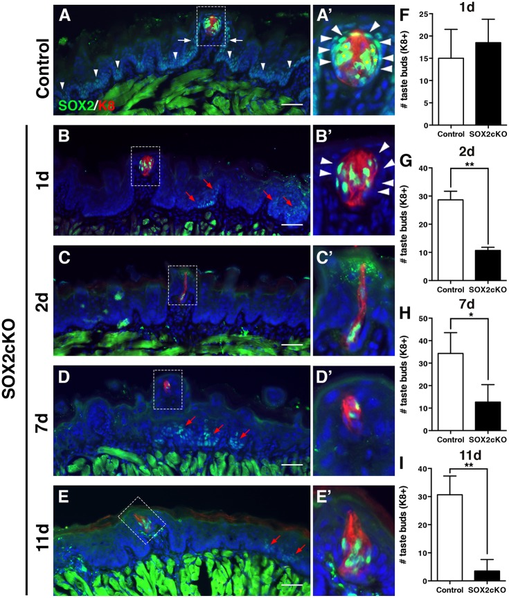
Genetic ablation of Sox2 in K14+ progenitors disrupts taste bud renewal. (A,A′) In control mice (K14+/+;Sox2flox/flox), SOX2 immunoreactivity (green) is high in taste bud cells (K8+, red, asterisks) and PG cells (A′, arrowheads). SOX2 is expressed at low levels by basal cells in FFP walls (white arrows) and non-taste epithelium (arrowheads in A). (B,B′) One day after Sox2 deletion (K14CreERT2/+;Sox2flox/flox), SOX2+ cells are found within most taste buds, PG cells lack SOX2 expression (B′, arrowheads) and SOX2+ epithelial cells outside of buds are limited to sparse, scattered clusters (B, red arrows). (C-E′) A similar pattern of SOX2 immunoreactivity is observed at 2 (C,C′), 7 (D,D′) and 11 (E,E′) days after Cre induction; SOX2+ cells are observed in occasional taste buds and scattered small clusters of more dimly SOX2+ cells are evident in non-taste epithelium (red arrows). A′-E′ show high magnification views of the boxed areas in A-E, respectively. (F) Taste bud number in mutant mice does not differ from controls 1 day after Sox2 deletion. (G) Deletion of Sox2 results in significant loss of K8+ taste buds after 2 days. (H) A similar reduction in taste bud number is evident at 7 days post-SOX2cKO, and by 11 days only 15% of taste buds remain in mutant tongues (I). The morphology of most remaining FF taste buds is disrupted in mutant mice; taste buds have fewer cells and/or more elongate morphology compared with controls (A′-E′). Nuclei are counterstained with Draq5 (blue). Scale bars: 50 μm. All are fluorescence images. N=3-5 mice per condition. Data are represented as mean+s.d.; *P<0.05, **P<0.01 (Student's t-test).

