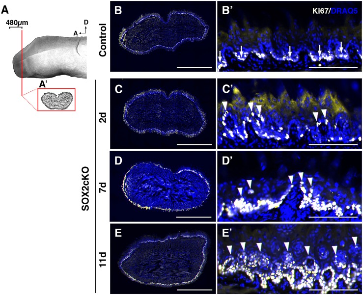
Fig. 6.
Proliferation is disorganized in SOX2cKO mice. (A) Proliferating cells (Ki67+) were assessed in representative transverse sections through the anterior tongue (A′) (first 480 μm from the tip). (B,B′) In controls, Ki67+ (light yellow) cells are restricted to the basal layer of the lingual epithelium. (C-E′) In SOX2cKO mice, Ki67+ cells also reside basally, but progressively more Ki67+ cells are found in suprabasal layers at later times post SOX2cKO (C′-E′, arrowheads). B′-E′ show high magnification views of the dorsal lingual epithelium in B-E, respectively. Nuclei are counterstained with Draq5 (blue). All images are scanned best focus sections. A, anterior; D, dorsal. Scale bars: 1 mm (B-E); 125 μm (B′-E′).

