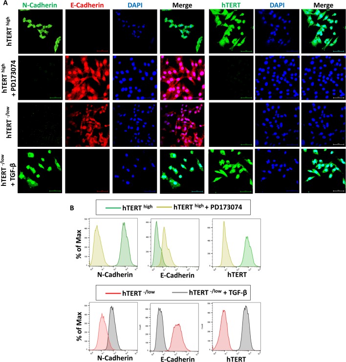
Fig. 3.
hTERT expression in CSCs is mutually exclusive with the mesenchymal phenotype. (A) Confocal immunofluorescence images for N-cadherin (green), E-cadherin (red) and Snail+Slug (green) showing that the loss of mesenchymal phenotype in hTERThigh CSCs mediated by PD173074 is associated with the loss of hTERT expression. However, acquisition of a mesenchymal phenotype in hTERT-/low CSCs mediated by TGF-β is associated with increased hTERT expression. Nuclei were stained with DAPI (blue). Scale bars: 60 µM. (B) Flow cytometry overlay histogram analysis of N-cadherin, E-cadherin and hTERT showing that hTERThigh CSCs treated with PD173074 lose their mesenchymal phenotype, which was associated with a loss of hTERT expression. Additionally, hTERT-/low CSCs treated with TGF-β acquired a mesenchymal phenotype, which was associated with increased hTERT expression. For comparison, an isotype control was used to define the positive and negative population for each marker.

