Fig. 4.
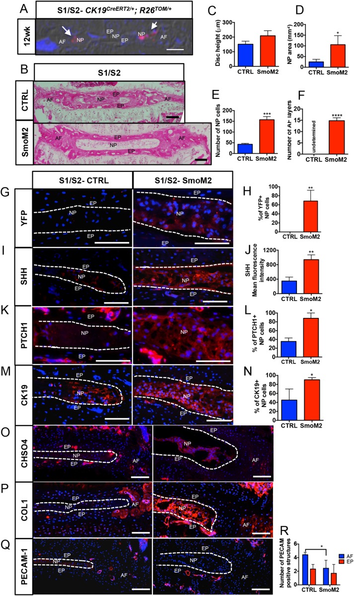
Constitutive activation of SmoM2 in the NP cells re-activates the sacral disc. (A) Coronal section of S1/S2 discs from CK19CreERT2/+; R26TOM/+ gavaged at 11 weeks of age and analyzed 1 week later. Red cells are the CK19CreERT2/+ expressing cells that have undergone recombination (white arrows). (B) H and E staining of the mid-coronal section from the control (CTRL- R26SmoM2-YFP/SmoM2-YFP) and SmoM2 (CK19CreERT2/+; R26SmoM2-YFP/SmoM2-YFP) littermates. (C-F) Quantification of morphometric parameters disc height (C); area occupied by the NP cells (D); the number of NP cells (E); and the number of layers in the AF (F) in the sacral discs of the SmoM2 group compared to littermate controls. (G,H) Immunostaining and quantification of YFP+ NP cells in SmoM2 and control discs. (I-P) Data from immunostaining for SHH and its downstream targets and their quantification: SHH (I,J); PTCH1 (K,L); CK19 (M,N); CHSO4 (O); COL1 expression in S1/S2 discs from controls and SmoM2 mice (P). (Q,R) Immunostaining and quantification of PECAM-1 positive structures in the AF and EP of control and SmoM2 mouse discs. Mean±s.d. N=4 Controls; N=3 SmoM2. Unpaired two-tailed t-test. *P<0.05, **P<0.01, ***P<0.001, ****P<0.0001. Scale bars: 100 μm. Nuclei are counter-stained with DAPI in A, G, I, K, M, O, P and Q. A is captured using DIC filter.

