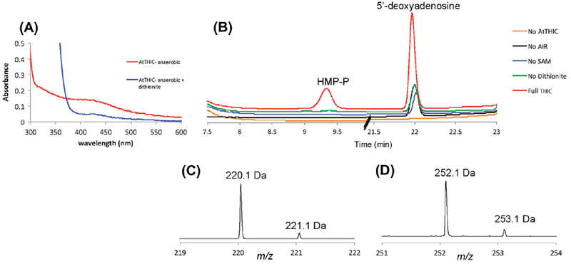
Fig.1.

ΔN71-AtTHIC activity measurement. (A) The red trace corresponds to anaerobically purified ΔN71–AtTHIC showing a UV–visible spectrum with a maximum at 410 mm and a shoulder at 320 nm, characteristic of a [4Fe–4S] cluster. Upon addition of dithionite, reduction of the cluster leads to the UV–visible spectrum shown in the blue trace with a characteristic absorption maximum at 425 nm. (B) HPLC profiles for the formation of HMP-P and 5ʹ-deoxyadenosine as measured by the absorbance at 254 nm. The red trace clearly indicates that the HMP-P is formed (elution time 9.4 min) only in assays where ΔN71–AtTHIC, AIR, SAM, and dithionite are present (All). No HMP-P formation is detected in controls where eitherAIR, SAM, ΔN71–AtTHIC or dithionite is absent. High amounts of 5ʹ-deoxyadenosine formation are seen in assays where ΔN71–AtTHIC, AIR, SAM, and dithionite are present (All). Loweramounts of 5ʹ-deoxyadenosine (elution time 22 min) are seen in assays where eitherAIR, SAM ordithionite is absent and no 5ʹ-deoxyadenosine is detected in assays where ΔN71–AtTHIC is absent. This suggests that a small amount of 5ʹ-deoxyadenosine co-purifies with ΔΝ71–AtTHIC. (C and D) ESI-MS (positive mode) of the reaction products eluting at 9.4 min (C, HMP-P) and 22 min (D, 5ʹ-deoxyadenosine).
