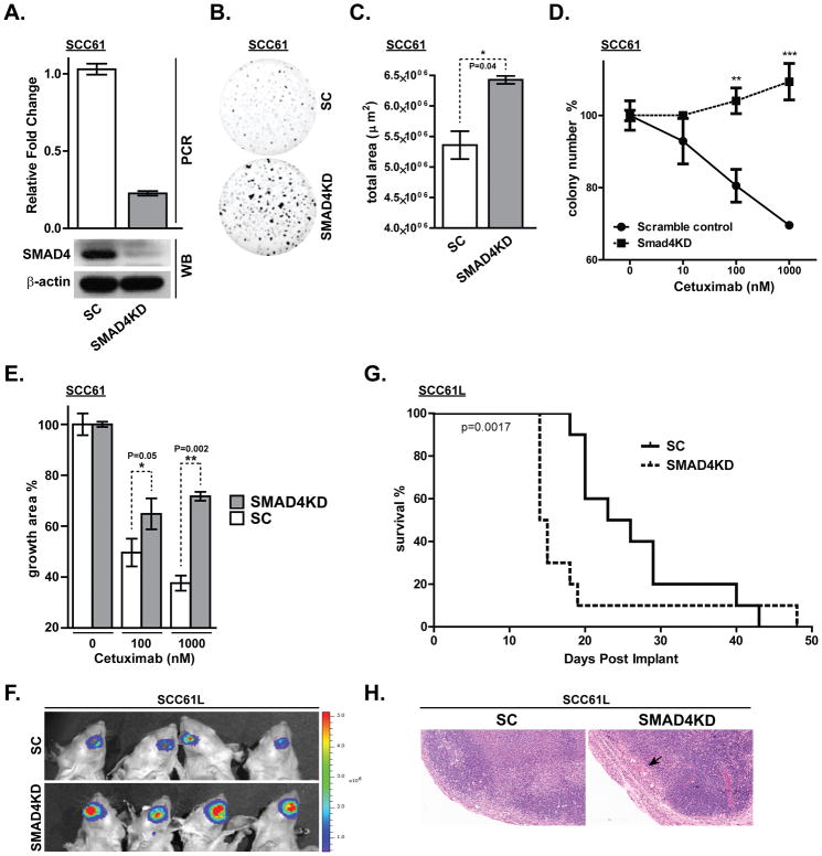
Figure 2. SMAD4 depletion in an HPV (−) HNSCC cell line increases clonogenic survival and cetuximab resistance.
(A) Real time PCR and western blot (WB) confirming knockdown of SMAD4 expression by stable expression of short-hairpin RNA in the SCC61 cell line. (SC: scrambled control shRNA, SMAD4KD: shRNA against SMAD4). (B, C) Clonogenic survival was determined by Matrigel colony formation assay for 7 days and total area was quantified (*p<0.05). Matrigel colony formation assay was performed for 7 days in PBS, 10nM, 100nM or 1000nM cetuximab showing (D) colony number (**p<0.01. ***p<0.0001) and (E) percent growth area. (F) SCC61L-SC and SCC61L-SMAD4KD cells that stably expresses firefly luciferase were submucosally injected into oral tongue of athymic nude mice. Bioluminescence images were taken at 13 days after tumor cell inoculation. (G) Kaplan-Meier survival curves for orthotopic xenograft mouse models of SCC61L-SC and SCC61L-SMAD4KD cells. (H) Representative H&E staining of tumor free lymph node derived from SCC61L-SC xenograft model and lymph node harvested from SCC61L-SMAD4KD model that shows metastatic tumor nodules (black arrow). Bar, 200 μm.

