Figure 3.
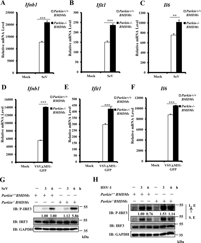
Parkin knockout potentiates type I IFN response in macrophage cells. A–C, Parkin deficiency significantly enhanced SeV-induced transcript levels of antiviral genes in BMDMs. Parkin+/+ and Parkin−/− BMDMs were infected with SeV for 6 h and then lysed for qRT–PCR analysis to examine transcript levels of Ifnb1 (A), Ifit1 (B), and Il6 (C). D–F, Parkin deficiency enhanced VSVΔM51-GFP-induced transcript levels of antiviral genes in BMDMs. Parkin+/+ and Parkin−/− BMDMs were infected with VSVΔM51-GFP virus at MOI of 1 for 6 h and then lysed for qRT–PCR analysis to measure transcript levels of Ifnb1 (D), Ifit1 (E), and Il6 (F). G, SeV-induced IRF3 phosphorylation was increased in Parkin−/− BMDMs. Parkin+/+ and Parkin−/− BMDMs were left untreated or infected with SeV for the indicated times. The cell lysates were separated by SDS–PAGE and subjected to immunoblotting (IB) with the indicated antibodies. H, HSV-1–induced IRF3 phosphorylation was enhanced in Parkin−/− BMDMs. Parkin+/+ and Parkin−/− BMDMs were left untreated or infected with HSV-1 at MOI of 5 for the indicated times. The cell lysates were separated by SDS–PAGE, followed by immunoblotting with the indicated antibodies. The data in A–F are from a representative experiment of at least three independent experiments (means ± S.D. of triplicate experiments). Two-tailed Student's t test was used to determine statistical significance. **, p < 0.01; ***, p < 0.001, versus control groups. Numbers below lanes (top) in G and H indicate densitometry of the protein presented relative to IRF3 expression in the same lane. S. E, short exposure; L. E, long exposure.

