
Figure 2.
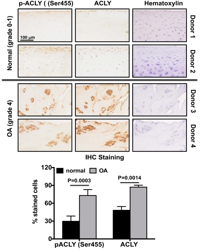
ACLY was up-regulated in human knee OA cartilage in situ. Human knee cartilage serial sections were analyzed for ACLY phosphorylation (Ser-455) and expression by immunohistochemistry. Hematoxylin staining verified cellularity in each section. Data are representative of 2 normal donors (24- and 45-year-old male) and 2 OA donors (84- and 90-year-old female) from 8 different donors (4 normal 4 OA) studied. The graph represents percentage of cells stained positively for phosphorylated ACLY (Ser-455) or ACLY. Student's t test was used for statistical data analysis. p values represent comparisons of the mean ± S.D.