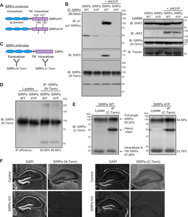
Figure 1.
Characterization of SIRPα mutant constructs and antibodies. A, illustration of the structures of SIRPαWT and SIRPα4YF. Four intracellular tyrosine residues in SIRPαWT were replaced with phenylalanine to generate SIRPα4YF. TM, transmembrane domain. B, verification of the tyrosine phosphorylation-deficient mutant of SIRPα (SIRPα4YF). COS cells were transfected with a SIRPα construct (WT or 4YF) and SHP2, together with or without an active form of JAK2 (JAK2VF). Cells were immunoprecipitated for SIRPα protein (with SIRPα N-Term antibody; described below), and the immunoprecipitates were blotted for phosphotyrosine (pY), SHP2, and SIRPα (SIRPα N-Term) (left). SIRPαWT is phosphorylated by JAK2VF and binds SHP2; however, SIRPα4YF is not phosphorylated by JAK2VF and does not bind SHP2. Expression levels of transfected proteins in the lysates are shown in the right panel. C, illustration showing the recognition sites of the anti-SIRPα antibodies used; SIRPα (N-Term) recognizes the SIRPα ectodomain, and SIRPα (C-Term) recognizes the SIRPα intracellular domain. D, immunoprecipitation efficiency of the SIRPα (N-Term) antibody. The same amount of lysates was used for direct blotting (Lysates) and immunoprecipitation (IP) followed by blotting. IP efficiency (%) was calculated by quantifying the ratio of SIRPα (IP) to SIRPα (Lysates). The IP efficiency of the SIRPα (N-Term) antibody in detecting SIRPαWT and SIRPα4YF was 83.88 and 86.66%, respectively. E, immunoprecipitation efficiency of the SIRPα (C-Term) antibody. The C-Term antibody recognizes both the full-length and the intracellular/transmembrane domain (lacking the ectodomain) SIRPα. IP efficiency was calculated as in D. The SIRPα (C-Term) effectively immunoprecipitated SIRPαWT (full-length, 95.93%; intracellular fragment, 87.89%) and SIRPα4YF (full-length, 94.58%; intracellular fragment, 93.78%). F, verification of the specificity of SIRPα antibodies by immunostaining. SIRPα KO brains were stained with either SIRPα (N-Term) or SIRPα (C-Term). SIRPα KO brains showed a lack of staining with both antibodies.

