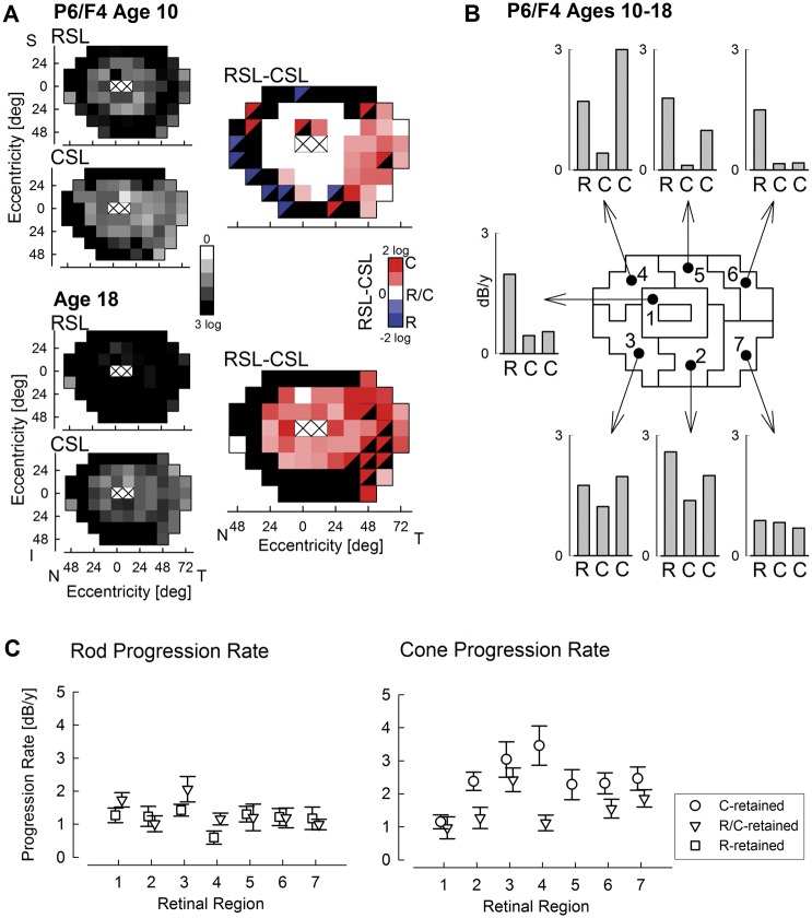
Figure 5.
Long-term progression of rod and cone dysfunction. (A) Rod- and cone-mediated sensitivity losses (RSL and CSL, smaller greyscale maps) and classification of whether rod, cone or both photoreceptors are retained (same as Fig. 4) in a representative patient recorded at ages 10 and 18 show substantial and non-homogeneous progression. (B) Calculated progression rates in dB per year across the retina tiled into seven neighbouring regions. Progression rates for rods and cones are specified for those loci that were R/C-retained at first visit (R and centre C, respectively), and for those loci that were C-retained at first visit (right C). Progression rates for loci with R-retention at first visit were not calculated since rod function at most of these points was undetectable at the second visit. (C) Rod and cone function progression rates across all patients with long-term followup as a function of retinal region described in Panel B. Rod progression is specified at loci with R- and R/C-retention at first visit, and cone progression at loci with C- and R/C-retention at the first visit.

