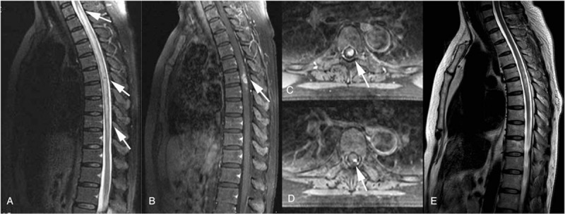
Figure 1.

(A) A sagittal T2-weighted image of the thoracic spinal cord shows a long segment of diffuse high-signal intensity in the central portion of the spinal cord from T1 to T10. (B) A sagittal contrast-enhanced T1-weighted image shows enhancement of the pia mater at the T6 level. The superficial parts of T6 also strongly enhanced with gadolinium and showed a candle guttering appearance. The abnormal parenchymal enhancement was relatively reduced on T2-weighted images, which showed the characteristic “flip-flop sign.” (C, D) Axial T1-weighted images with contrast enhancement at the T6 level. (E) After treatment with ceftriaxone, the thoracic lesion has diminished, and might represent meningeal inflammation and spinal cord ischemia or edema.
