Figure 2.

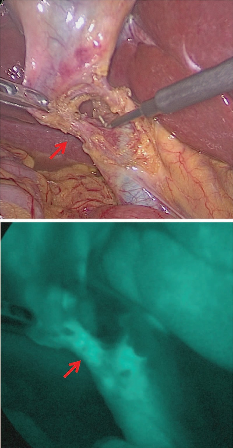
Upper panel was detected by a “white” light source and cystic duct was detected in lower panel by a “near infrared” light source after dissecting Calot triangle.

Upper panel was detected by a “white” light source and cystic duct was detected in lower panel by a “near infrared” light source after dissecting Calot triangle.