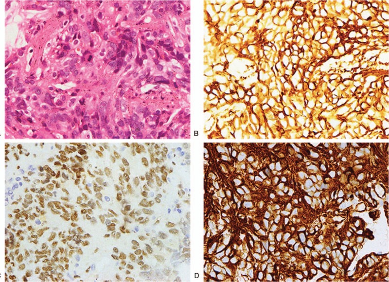
Figure 4.

Epithelioid angiosarcoma was identified by H&E and immunohistochemical staining. (A) H&E staining (×400). (B) High expression of CD 31 by immunohistochemical staining (×400). (C) High expression of ERG by immunohistochemical staining (×400). (D) High expression of VIM by immunohistochemical staining (×400).
