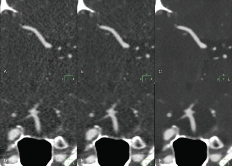
Figure 4.

Transverse CT images of the right middle cerebral artery (A–C) and coronal CT images of the basilar artery (D–F) reconstructed with FBP (A, D); hybrid IR (B, E); and IMR (C, F) at 80 kVp. Images reconstructed with IMR offer significant noise reduction and smoother vascular structure compared with FBP and hybrid IR. CT = computed tomography, FBP = filtered back projection, IMR = iterative model reconstruction, IR = iterative reconstruction.
