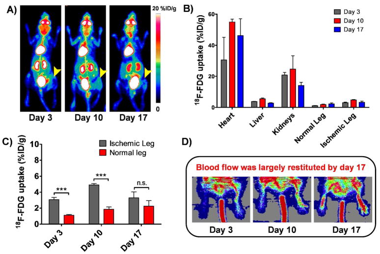
Figure 2.
A) Serial maximum intensity projection (MIP) PET images of mice injected with 18F-FDG on postoperative days 3, 10 and 17. Arrowhead points to the ischemic leg. Tracer uptake in the ischemic leg, despite being significantly higher in the ischemic leg, was still considered to be low. B) PET imaging-derived 18F-FDG uptake, reported as %ID/g ± SD, in different tissues on postoperative days 3, 10 and 17 (n=3). C) Comparative graph showing 18F-FDG uptake in the ischemic and non-operated legs on postoperative days 3, 10 and 17. D) Serial Laser Doppler imaging of non-ischemic (right side) and ischemic hindlimbs (left side) of balb/c mice undergone surgery. Dark blue areas, observed at day 3, indicate decreased perfusion in the injured limb, while high perfusion pattern (red color) is observed in the contralateral hindlimb. Some perfusion recovery was detectable by day 10 and blood flow was largely restituted in the operated hindlimb by day 17.

