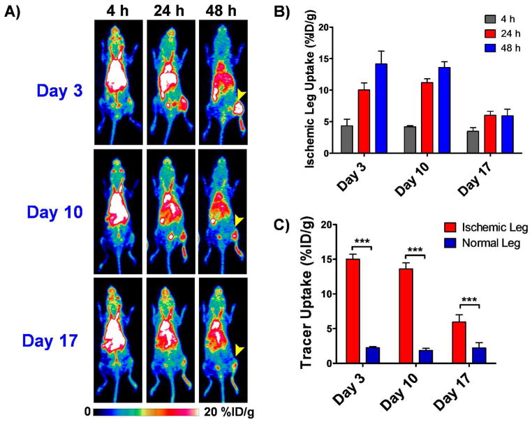
Figure 3.
Longitudinal PET imaging studies in ischemic mice injected intravenously with 5–10MBq (135–270 μCi) of 64Cu-NOTA-YY146 on postoperative days 3, 10 and 17. A) Serial maximum intensity projection (MIP) PET images at 4, 24 and 48h p.i. at the different postoperative timepoints. Arrow heads point to the ischemic leg. B) Tracer uptake, represented as mean %ID/g ± SD, in the ischemic leg at different time points on the different postoperative days. C) Comparative graph showing peak tracer uptake in the ischemic and non ischemic legs at 48h p.i. on postoperative days 3, 10 and 17.

