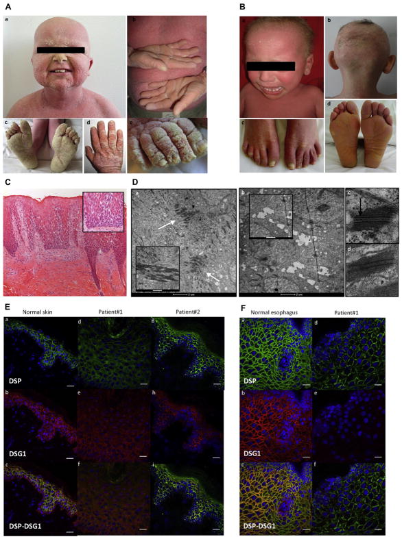
FIG 1.
Clinical and histopathologic features of patients 1 and 2. A, Clinical phenotype of patient 1: desquamative erythroderma, thickening of the skin (a–e), sparse hair (a), diffuse palmoplantar keratoderma (PPK; b and c), and thickening of the nail plate (d and e). B, Clinical phenotype of patient 2: desquamative erythroderma (a), sparse hair (a and b), onycholysis (c), and plantar keratoderma (d). C, Skin histology (patient 1) showing epidermal acanthosis, hyperorthokeratosis, extensive acantholysis (inset), and inflammation in the superficial dermis (lymphocytes; ×100 magnification for Fig 1, C, and ×200 magnification for the inset of Fig 1, C). D, Ultrastructural features of a skin biopsy specimen from patient 1 (a, b, and d) and a healthy control subject (c). a, Many desmosomes clustered in the upper epidermis (arrows). Keratin filaments strongly aggregated to the desmosome (inset). b, Complete absence of desmosomes and widened spaces between keratinocytes (cell membrane features filipodium-like processes; inset) in the lower epidermis. d, Patient 1’s desmosome lacks the inner plaque compared with a control subject (arrow, c). Scale bar = 2 μm for Fig 1, D, a and b. Scale bar = 500 nm for the inset of Fig 1, D, a, and 1 μm for the inset of Fig 1, D, b. E, Immunofluorescence on skin sections from a healthy control subject (a–c), patient 1 (d–f), and patient 2 (g–i) showed drastic reduction and mislocalization in staining of DSP and DSG1 in both patients. Scale bar =20 μM. F, Immunofluorescence on esophageal sections from a healthy control subject (a–c) and patient 1 (d–f), showing low levels of DSP staining and absence of DSG1 staining in patient 1. Scale bar = 20 μM.

