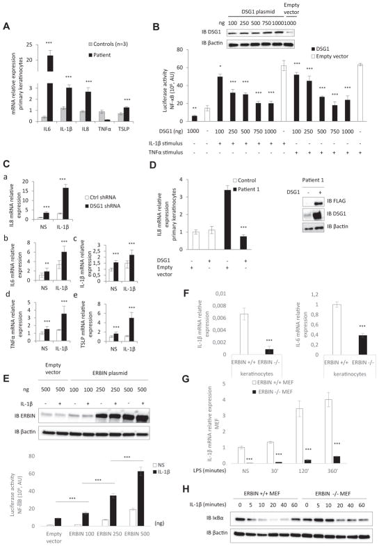
FIG 2.
Role of DSG1 and ERBIN in NF-κB–mediated epithelial inflammation. A, Relative mRNA expression levels of proinflammatory cytokines in primary keratinocytes (patient 1). B, NF-κB luciferase reporter assay in HEK293T cells transfected with DSG1 vector or with an empty vector and stimulated with IL-1β or TNF-α. AU, Arbitrary units. Inset, Western blot of DSG1 expression in HEK293T cells transfected with DSG1 vector. C, Relative mRNA expression of IL8 (a), IL6 (b), IL1B (c), TNFA (d), and TSLP (e) in control (ctrl) keratinocytes infected with a lentivirus-expressing short hairpin RNA against DSG1 and stimulated or not stimulated (NS) with IL-1β. D, Relative mRNA expression of patient 1’s keratinocytes infected with a retrovirus expressing FLAG DSG1. Inset, Western blot of DSG1 and FLAG expression in patient 1’s keratinocytes after transduction. E, NF-κB luciferase reporter assay in HEK293T cells transfected with ERBIN or empty vector and stimulated with IL-1β. Inset, Western blot of ERBIN expression in HEK293T cells transfected with the ERBIN vector. F, Relative mRNA expression levels of IL1B and IL6 in nonstimulated Erbin+/+ and Erbin−/− keratinocytes. G, Relative mRNA expression levels of IL1B in Erbin+/+ and Erbin−/− MEFs before/after stimulation with LPS. H, Immunoblot assays showing degradation of IκBα proteins in Erbin+/+ and Erbin−/− MEFs induced by IL-1β stimulation. *P < .05, **P < .01, and ***P < .001.

