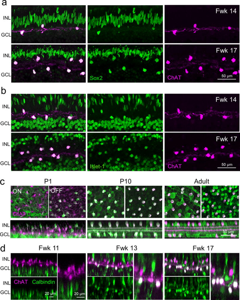
Figure 6. Molecular expression patterns of ON and OFF ChAT cells.

ChAT immunostaining together with immunolabeling for (a) Sox2 and (b) Islet-1 in the peri-fovea of the human retina at fetal weeks (Fwks) 14 and 17. (c) Calbindin and ChAT immunostaining in the postnatal day (P) 1, 9 and adult mouse retina. (d) Calbindin and ChAT immunostaining in the putative human fovea at different fetal ages. For each age, representative ON and OFF ChAT cells are displayed at higher magnification in the right panels. INL, inner nuclear layer; GCL, ganglion cell layer.
