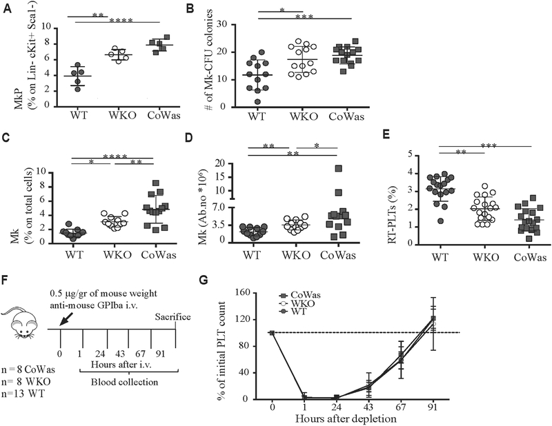
FIG 2.
Phenotypic and functional characterization of the megakaryocytic compartment. A, Evaluation of megakaryocyte progenitors (MkP) identified as CD41+CD150+ cells gated on the Lin−c-Kit+Sca1− compartment in WT, WKO, and CoWas mice is shown. B, The number of MKcolony-forming units was counted with the microscope by using a ×4 objective. C and D, MKs (CD61+CD41+ cells) are expressed as percentages on total BM cells (Fig 2, C) or absolute numbers (Fig 2, D) and analyzed in the bone marrow of age-matched WT, WKO, and CoWas mice. E, The percentage of RT-PLTs is assessed by using Thiazole Orange (TO). F, Platelet depletion was performed by using intravenous injection of anti-mouse GPIba (Emfret Analytics) according to the scheme indicated in the figure. G, Platelet counts were monitored daily by means of blood collection and expressed as percentages of initial platelet counts at time 0 at different time points. In all graphs each dot represents a different mouse from 1 (Fig 2, A), 2 (Fig 2, G), 4 (Fig 2, E), or 6 (Fig 2, B-D) independent experiments. All the graphs report means ± SDs, and statistical analysis was performed with 1-way ANOVA (Fig 2, A-E) or 2-way ANOVA (Fig 2, G) and the Bonferroni postcorrection test. *P < .05, **P < .005, ***P <.001, and ****P < .0001. PLT, Platelets.

