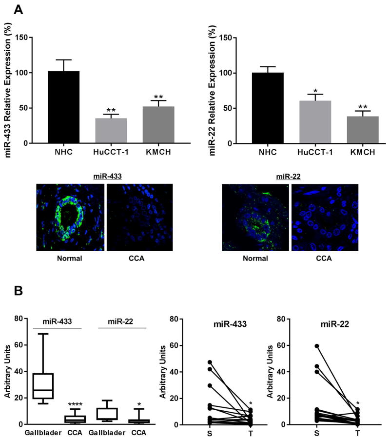
Figure 1. MicroRNAs targeting HDAC6 are downregulated in CCA.
A, The expression pattern of miR-433 and miR-22 was analyzed in normal cholangiocytes (NHC) and the CCA cell lines HuCCT-1 and KMCH by q-PCR. In situ hybridization for miR-433 and miR-22 in human cholangiocarcinoma tissues. Green, positive signals; blue, counterstained nuclei with DAPI. B, qPCR for miR-433 and miR-22 in a different cohort of CCA human samples (T) compared to matched surrounding tissue (S) and to normal gallbladders as controls. (*p<0.05, **** p<0.001)

