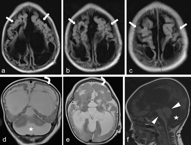
Fig. 3.
Axial fluid-attenuated inversion recovery (A-C), T2-weighted coronal (D) and axial (E), and T1-weighted sagittal (F) sequence of magnetic resonance images show polymicrogyria (arrows), agyri (curved arrows), brainstem-cerebellar atrophy (arrowheads), and enlargement of the retrocerebellar cyst (stars).

