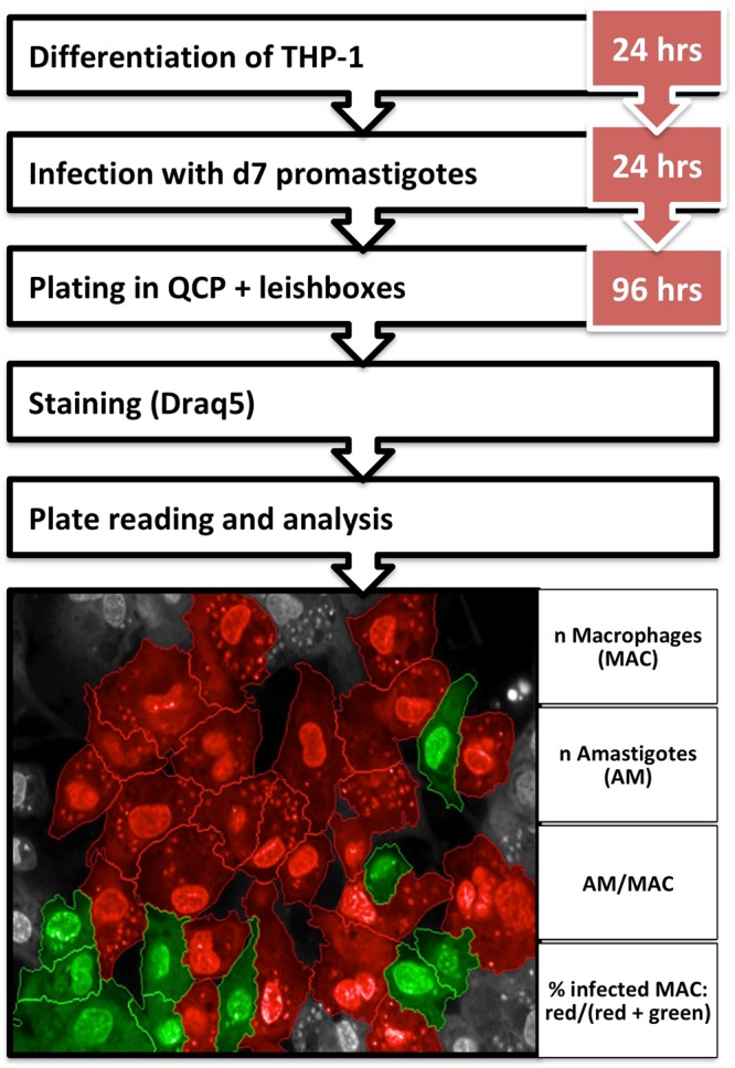
Figure 1.

Experimental setup and analysis parameters of the InMAC assay. THP-1 cells are differentiated for 24 hrs followed by o/n infection with d7 promastigotes. Infected THP-1 cells are plated and incubated with the “Leishbox” compounds and controls for 96 hrs. Finally plates are stained with Draq5, read and analysed. Images were taken by Opera at 40x magnification. The infected cells are shown in red while the non-infected ones are shown in green.
