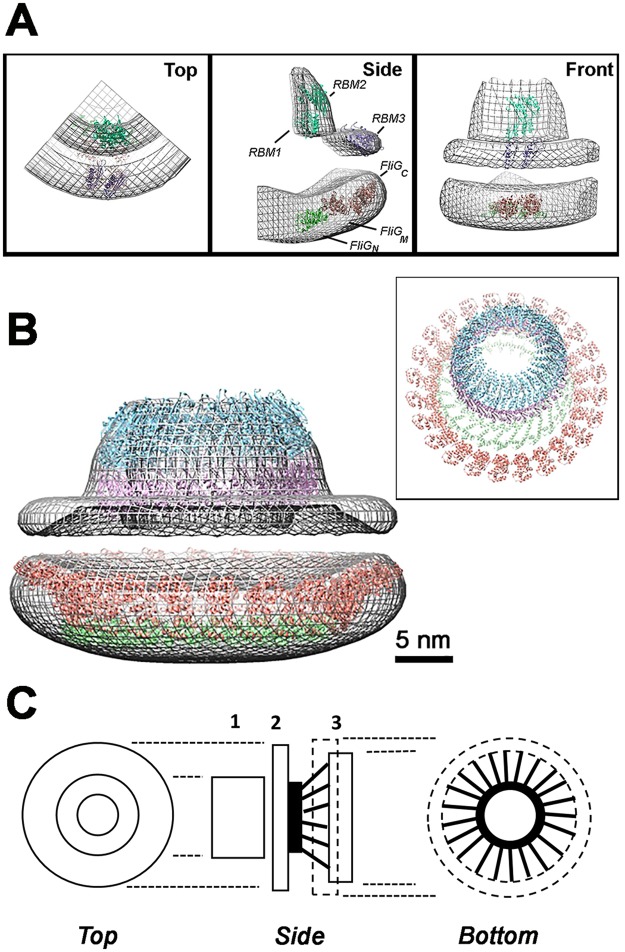
Figure 5.
Homodimer fits of FliF and FliG domains in the 3D reconstruction suggest a mechanical model. (A) CHIMERA61 best-fits of the EscJ dimer (RBM1 + RBM2; c = 0.87), split RBM3 (c = 0.83), T. maritima FliG_N (5TDY-A, B; c = 0.81) and FliG_MC (4FHR; 0.83) into the cylindrically-averaged 3D-reconstruction (mesh representation, 0.4 contour level, c = correlation coefficient). Fits were also made for the alternative T. maritima (5TDY-D; c = 0.80) and A. aeolicus (PDB ID = 3HJL; c = 0.69) FliG_N conformations. The sub-volumes were fit sequentially; first the EscJ dimer followed by the RBM3 dimer model for the periplasmic sub-volumes; the T. maritima FliG_N followed by the FliG_MC structures for the cytoplasmic sub-volume. Manual refinement improved correlation, albeit to a small degree, of the automated assignments. (B) CHIMERA sequential fit of 13 dimer rings of 1YJ7, RBM3 and FliG_N plus 26 FliG_MC in the reconstruction (0.42 electron density/voxel threshold, volume = (3.81*(10^6)) cubic angstroms). Box Inset. Oblique view of the sequential fit ring models without the electron density map. Atomic structures in B are color coded as in A. (C) Engineer’s sketch of the mechanical design of the FliFFliG fusion rotor. 1 = RBM1/RBM2 ring. 2 = RBM3 ring. 3 = FliG-N (solid)/FliG-MC (dashed) rings. FliF transmembrane (black ring) and cytoplasmic C-terminal helices (black rods).

