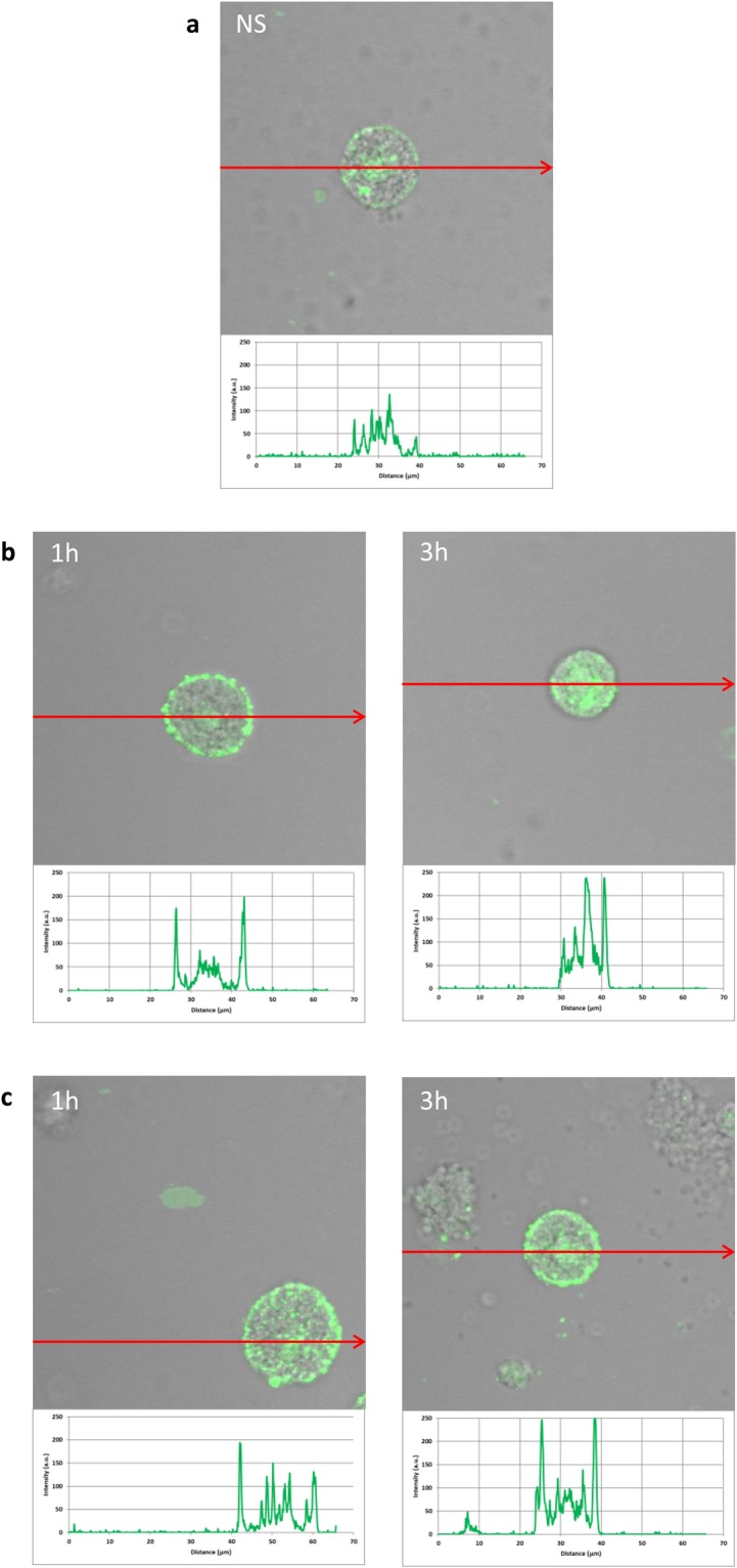
Figure 5.

Effect of LL-37 and hBD-2 stimulation on RIG-I expression in PMCs. PMCs were incubated with medium alone (non-stimulated cells; NS), LL-37 or hBD-2 at a final concentration of 1 µg/mL. Representative images showing RIG-I cellular localization in permeabilized (a) native, (b) stimulated LL-37, (c) stimulated hBD-2 PMCs analyzed by confocal microscopy. Single confocal sections (midsection of cells) reveal presence of RIG-I. One representative of three independent experiments performed with duplicate samples (each experiment was performed on 4 animals/PMC isolates) is shown. Fluorescence intensity diagrams showing the distribution of fluorescence in cells were mounted.
