Neutrophils are short-lived cells that can undergo many types of cell death
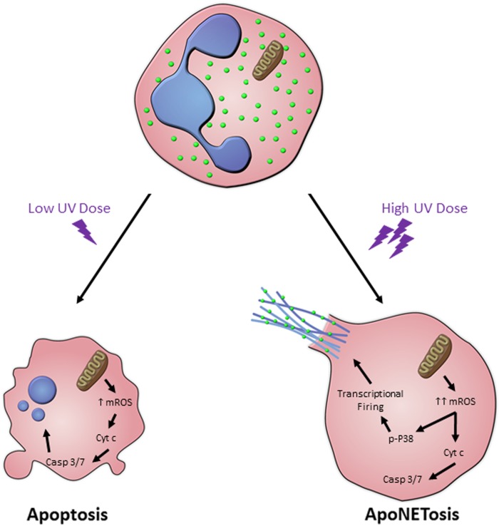
Neutrophils are the most abundant leukocyte in the human body. They have a short lifespan (average of 7 hours to 5.4 days) and types of neutrophil death affect many inflammatory and autoimmune disease states. Neutrophils have classically been known to die by apoptosis. A novel form of neutrophil death leading to the release of extracellular traps (NETs) that is different from apoptosis was discovered in 19961. In the past decade, mechanistic details of NET formation (NETosis) have been studied in greater detail2,3. NETosis has now been defined as a unique form of cell death that is completely independent from apoptosis1,2. We have recently reported that higher doses of ultraviolet (UV) induce both apoptosis and NETosis in the same neutrophil, in Cell Death Discovery4. In this novel form of neutrophil death, ApoNETosis, NETotic steps override certain key apoptotic steps (Fig. 1). ApoNETosis, apoptosis, NETosis, and other forms of neutrophil death have unique similarities and differences, and understanding molecular steps regulating ApoNETosis could help to explain certain pathobiological conditions.
Fig. 1. The key steps of UV-induced apoptosis and ApoNETosis.
Low dose UV irradiation induces low levels of mitochondrial ROS, cytochrome c release, caspase cascade activation, and finally nuclear condensation and blebbing. On the other hand, high dose UV irradiation results in large amounts of mitochondrial ROS, cytochrome c release, and caspase cascade activation. However, p38 activation and transcriptional firing are also induced, resulting in MPO-coated NET formation and NET release.
Apoptosis and necrosis are the classical forms of neutrophil death
There are two types of neutrophil apoptosis: intrinsic and extrinsic apoptosis. Intrinsic apoptosis is induced primarily by UV light and bacteria5,6. Following exposure to UV, mitochondrial membrane is destabilized resulting in the release of cytochrome c7. Once in the cytoplasm, cytochrome c binds the adapter protein Apaf1. The complex activates pro-caspase 9 to form caspase 9. Caspase 9 then activates the effector proteins such as caspase 3/7, which then activates caspase-activated DNase (CAD)7. Extrinsic apoptosis is induced primarily by Fas ligand and tumor necrosis factor α (TNFα)8. Fas and tumor necrosis factor receptor-1 (TNFR-1) are membrane proteins that are activated by Fas ligand and TNFα, respectively8. Activation of either membrane protein results in the formation of the death-inducing signaling complex (DISC) composed of Fas, Fas-associated protein with Death Domain (FADD), and pro-caspase 8. This results in the activation of caspase 8, which in turn activates effector caspases 3/7 followed by the activation of CAD8. Depending on phosphorylation state, caspase 8 has also been shown to prolong survival of neutrophils6. Bcl-2 family members play important roles in neutrophil apoptosis. Neutrophils express high levels of pro-apoptotic Bcl-2 family members, Bax and Bak, which may play a role in their observed short lifespans9. Pro-apoptotic Bid, Bim, and Bad are also expressed in neutrophils9. Anti-apoptotic Bcl-2 family members, Mcl-1, Bcl-xl, and A1, are expressed in neutrophils and may be increased by survival factors9.
There are many factors and signaling pathways that affect neutrophil apoptosis. granulocyte/macrophage colony-stimulating factor, granulocyte colony-stimulating factor, and ATP are a few of the factors that promote neutrophil survival9. On the other hand, activation of certain tumor necrosis factor/nerve growth factor receptor family members can result in enhanced apoptosis9.
While apoptosis is highly regulated, necrosis can be uncontrolled and often accidental. It is often the result of stresses such as osmotic shock, heat and freeze thawing. Higher levels of bacteria have also been reported to induce necrosis in neutrophils. There is a regulated type of necrosis termed necroptosis, which can be induced by bacterial pathogen-associated molecular patterns and viral infections. Necroptosis is dependent on RIPK1 and RIPK3 activation and negatively dependent on caspase 810.
NETosis is a cell death that is specific to neutrophils
A form of cell death specific to neutrophils is NETosis. NETosis, which is independent of apoptosis and necrosis, was first observed by Takei et al in 19961. The research team induced NETosis by exposing the neutrophils to PMA1. The bactericidal activity of NETs was uncovered by Brinkmann et al2. Since its discovery, two types of NETosis have been characterized3,11. The first type is NADPH oxidase 2 (NOX2)-dependent NETosis, which is induced by PMA, LPS, and various types of bacteria such as Pseudomonas aeruginosa. During NOX-dependent NETosis, ROS is produced by NOX2, mitogen-activated protein kinases (MAPKs: ERK, p38, and JNK) are activated, transcriptional firing is initiated and ultimately NETs are released11–13. The second type is NOX-independent NETosis, which is induced by calcium ionophores (A2317 and ionomycin) some crystals, certain microbes, and UV light4,11. During NOX-independent NETosis, increased ROS is produced by the mitochondria, MAPK p38 is activated, transcriptional firing is increased and ultimately NETs are released3,13. Specific to NETosis induced by calcium ionophores, intracellular calcium increase results in activation of peptidylarginine deiminase 4 (PAD4), which citrullinates histones at promoter regions that plays a role in accelerating the formation of NETs3,14. While NETosis has been classically seen as a type of cell death, a non-lytic form of NETosis has been uncovered and termed as vital NETosis15.
ApoNETosis: simultaneous apoptosis and NETosis in the same neutrophil
The classical thinking has been that NETosis and apoptosis are two distinct processes. In regards to the interaction between apoptosis and NETosis, caspase activity (a hallmark of apoptosis) was reported to be lacking during induction of NETosis1,2. No incidence of simultaneous induction of apoptosis and NETosis in the same neutrophil has been previously reported. This highlights the significance of the uncovering of the ability of UV, at higher doses, to induce NETosis and apoptosis simultaneously in the same neutrophil in a novel form of cell death (Fig. 1). ApoNETosis, similar to apoptosis and all types of NETosis, requires increased ROS production, specifically mitochondrial in origin in the case of ApoNETosis. NOX is not active during UV-induced ApoNETosis, indicating that it is NOX-independent. Activation of MAPK p38 is required for UV-induced ApoNETosis. Similar to other forms of NOX-independent NETosis, ApoNETosis requires transcriptional firing to take place4. However, it differs from other forms of NOX-independent NETosis in that histones are not citrullinated. This suggests that there is no increase in intracellular calcium concentration and PAD4 activation during UV exposure of neutrophils. Similar to other types of apoptosis, caspase 3 is cleaved during UV-induced ApoNETosis, indicating that apoptotic pathways are activated. However, nuclear blebbing does not occur during ApoNETosis. Instead DNA is released in NET structures as seen during NETosis.
Significance and future directions
NETosis is a form of cell death that is independent of apoptosis and necrosis. We uncovered that UV at higher doses can induce both apoptosis and NETosis in the same neutrophils in a novel process termed ApoNETosis. High dose UV as a tool for generating NETs for NET clearance studies is promising since drawbacks of using chemicals, toxins, or cytokines are not present in this experimental approach. The discovery that UV can induce ApoNETosis may shed light on states of inflammation observed following exposure to UV. Further mechanistic details and pathological relevance of ApoNETosis remain to be established.
Acknowledgements
This study was supported by research grants of the Canadian Institutes of Health Research (MOP-111012 to N.P.), Cystic Fibrosis Canada (Discovery Grant 3180 to N.P), and Natural Sciences and Engineering Research Council of Canada (RGPIN-2018-06575 to N.P.). D.A. is a recipient of an Ontario Student Opportunity Trust Fund/Restracomp studentship of SickKids.
Conflict of interest
The authors declare that they have no conflict of interest.
Footnotes
Publisher's note: Springer Nature remains neutral with regard to jurisdictional claims in published maps and institutional affiliations.
References
- 1.Takei H, Araki A, Watanabe H, Ichinose A, Sendo F. Rapid killing of human neutrophils by the potent activator phorbol 12-myristate 13-acetate (PMA) accompanied by changes different from typical apoptosis or necrosis. J. Leukoc. Biol. 1996;59:229–240. doi: 10.1002/jlb.59.2.229. [DOI] [PubMed] [Google Scholar]
- 2.Brinkmann V, et al. Neutrophil extracellular traps kill bacteria. Science. 2004;303:1532–1535. doi: 10.1126/science.1092385. [DOI] [PubMed] [Google Scholar]
- 3.Douda DN, Khan MA, Grasemann H, Palaniyar N. SK3 channel and mitochondrial ROS mediate NADPH oxidase-independent NETosis induced by calcium influx. Proc. Natl. Acad. Sci. 2015;112:2817–2822. doi: 10.1073/pnas.1414055112. [DOI] [PMC free article] [PubMed] [Google Scholar]
- 4.Azzouz D, Khan MA, Sweezey N, Palaniyar N. Two-in-one: UV radiation simultaneously induces apoptosis and NETosis. Cell Death Discov. 2018;4:51. doi: 10.1038/s41420-018-0048-3. [DOI] [PMC free article] [PubMed] [Google Scholar]
- 5.Frasch SC, et al. p38 mitogen-activated protein kinase-dependent and-independent intracellular signal transduction pathways leading to apoptosis in human neutrophils. J. Biol. Chem. 1998;273:8389–8397. doi: 10.1074/jbc.273.14.8389. [DOI] [PubMed] [Google Scholar]
- 6.Jia SH, Parodo J, Kapus A, Rotstein OD, Marshall JC. Dynamic regulation of neutrophil survival through tyrosine phosphorylation or dephosphorylation of caspase-8. J. Biol. Chem. 2008;283:5402–5413. doi: 10.1074/jbc.M706462200. [DOI] [PubMed] [Google Scholar]
- 7.Simon HU, Haj-Yehia A, Levi-Schaffer F. Role of reactive oxygen species (ROS) in apoptosis induction. Apoptosis. 2000;5:415–418. doi: 10.1023/A:1009616228304. [DOI] [PubMed] [Google Scholar]
- 8.Croker BA, et al. Fas-mediated neutrophil apoptosis is accelerated by Bid, Bak, and Bax and inhibited by Bcl-2 and Mcl-1. Proc. Natl. Acad. Sci. 2011;108:13135–13140. doi: 10.1073/pnas.1110358108. [DOI] [PMC free article] [PubMed] [Google Scholar]
- 9.Simon HU. Neutrophil apoptosis pathways and their modifications in inflammation. Immunol. Rev. 2003;193:101–110. doi: 10.1034/j.1600-065X.2003.00038.x. [DOI] [PubMed] [Google Scholar]
- 10.Kaczmarek A, Vandenabeele P, Krysko DV. Necroptosis: the release of damage-associated molecular patterns and its physiological relevance. Immunity. 2013;38:209–223. doi: 10.1016/j.immuni.2013.02.003. [DOI] [PubMed] [Google Scholar]
- 11.Douda DN, Yip L, Khan MA, Grasemann H, Palaniyar N. Akt is essential to induce NADPH-dependent NETosis and to switch the neutrophil death to apoptosis. Blood. 2014;123:597–600. doi: 10.1182/blood-2013-09-526707. [DOI] [PubMed] [Google Scholar]
- 12.Khan MA, et al. JNK activation turns on LPS-and gram-negative bacteria-induced NADPH oxidase-dependent suicidal NETosis. Sci. Rep. 2017;7:3409. doi: 10.1038/s41598-017-03257-z. [DOI] [PMC free article] [PubMed] [Google Scholar]
- 13.Khan MA, Palaniyar N. Transcriptional firing helps to drive NETosis. Sci. Rep. 2017;7:41749. doi: 10.1038/srep41749. [DOI] [PMC free article] [PubMed] [Google Scholar]
- 14.Rohrbach AS, Slade DJ, Thompson PR, Mowen KA. Activation of PAD4 in NET formation. Front. Immunol. 2012;3:360. doi: 10.3389/fimmu.2012.00360. [DOI] [PMC free article] [PubMed] [Google Scholar]
- 15.Yipp BG, Kubes P. NETosis: how vital is it?. Blood. 2013;122:2784–2794. doi: 10.1182/blood-2013-04-457671. [DOI] [PubMed] [Google Scholar]


