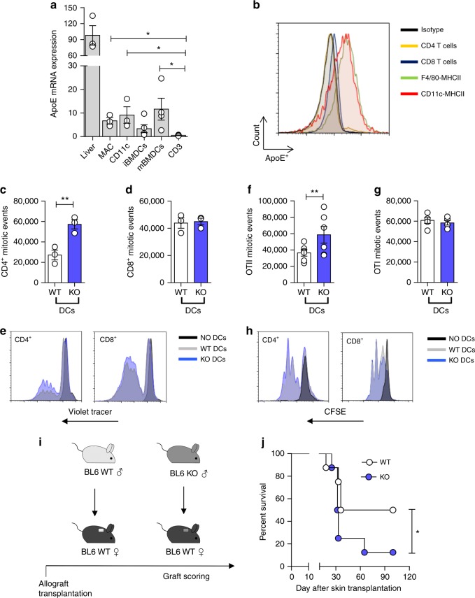
Fig. 3.
ApoE regulates DC activation. a mRNA expression relative to RPL (L Ribosomal Protein) of apoE in the liver, resident peritoneal macrophages (MAC), spleen-derived DCs (CD11c+), bone marrow derived DCs (immature iBMDCs and mature mBMDCs) and T cells (CD3+) of WT animals. b representative histogram showing apoE protein determination through flow cytometry in T cells (CD4+ and CD8+), macrophages (F4/80+ MHCII+) and DCs (CD11c+ MHCII+) of WT mice. c–e Proliferation of allogenic BALB/c CD4+ (c) and CD8+ (d) T cells with spleen-derived DCs from WT and apoE KO C57BL/6 mice; representative histograms are presented in e. f–h Proliferation of transgenic OTII CD4+ (f) and OTI CD8+ (g) T cells with BMDCs isolated from WT and apoE KO mice and pulsed with OTII or OTI peptide; representative histograms are presented in h. i Graphic representation of skin allograft transplantation: a piece of tail skin from a C57BL/6 male donor either WT or apoE KO was transplanted on the back of WT female mice; graft survival was scored up to 100 days. j Percentage of graft survival following skin transplantation. Graft survival of <50% of the donor skin was recorded as rejection. N = 3 (in triplicates c–d), N = 4 (a), N = 6 (f–g), N = 8 (j) per group. Statistical analysis was performed with unpaired T-test (a, c, d, f, g), Gehan-Breslow-Wilcoxon test (j). Data are reported as mean ± SEM; *p < 0.05, **p < 0.01

