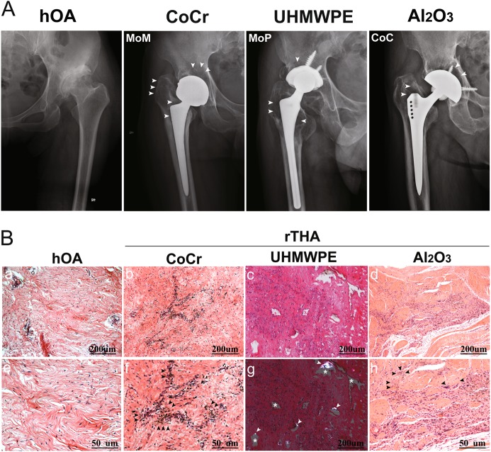
Fig. 1. Imaging and histomorphological manifestations of prosthesis loosening with different frictional interfaces.
a Anterior and posterior X-ray images demonstrating a wide range of osteolytic areas (white arrow) around the proximal femur and the acetabular cup at the MoM, MoP, and CoC interfaces in hOA patients. b (a, e) H&E staining showed that inflammatory cells infiltrated into the synovial tissue of patients with hOA. b, f H&E staining of the synovial tissue of the hip joint of patients with rTHA (metal-on-metal interface, MoM) showed a large number of black particles of uniform size (black arrow). c H&E staining of the synovial tissues from the hip joints of patients with rTHA (metal-on-polyethylene interface, MoP) showed that the uneven area of the blank (white asterisk) was surrounded by increased synovial fibroblasts and aggregated mononuclear macrophages in the synovium from patients with rTHA. (g) UHMWPE particles were observed as double refractions by polarized light micrographs (white arrow). d, h Gray or black micro- and nanosized scattered ceramic particles were observed in the synovium with fewer inflammatory cells (black arrow) from patients with rTHA (ceramic-on-ceramic interface, CoC). a, b, c, d, g Scale bar: 200 μm; (e, f, h) Scale bar: 50 μm. Revision total hip arthroplasty (rTHA), hip osteoarthritis (hOA); CoCr, UHMWPE, and Al2O3 refer to the wear debris of MoM, MoP, and CoC, respectively

