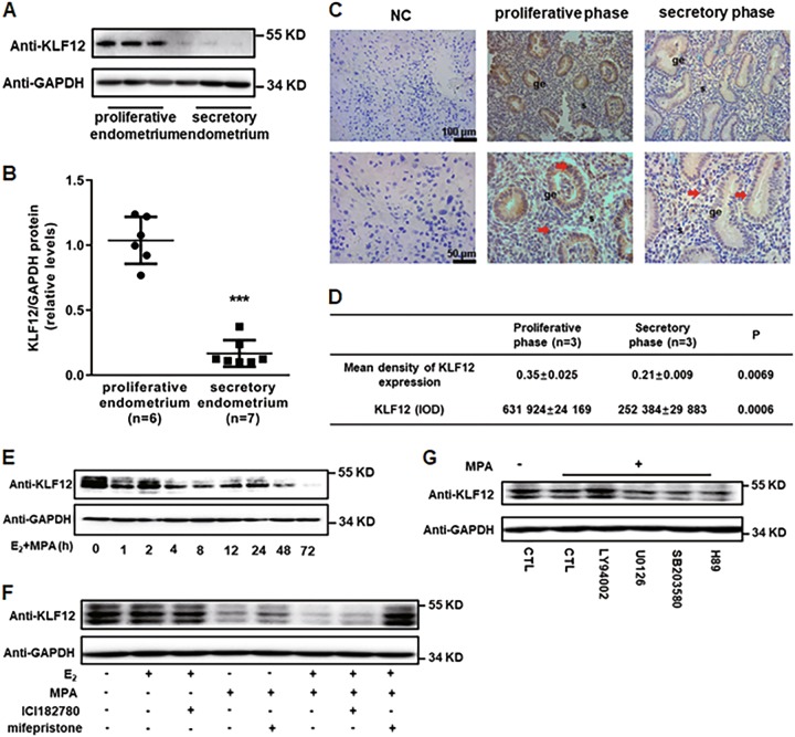
Fig. 3. KLF12 is a downstream gene that is regulated by progesterone.
a, b The total KLF12 protein levels of proliferative- and secretory-phase endometria were normalized to GAPDH expression, and the data for all of the endometrial samples are shown in the scatter plots. ***P < 0.001 compared with the proliferative endometrium. c, d Immunohistochemical analysis was performed using KLF12 antibody. Proliferative- and secretory-phase endometrial tissue samples from normal women are shown at ×200 (left panel) and ×400 (right panel) magnification. The negative control (NC) is nonspecific rabbit serum. Red represents positive staining (arrows). Scale bars, 100 μm (up panel) and 50 μm (below panel). The means and integrated optical densities of KLF12 expression in the endometrial tissue were calculated. e The Ishikawa cells were treated with estrogen (E2) (10–8 M) and progesterone (MPA) (10–6 M) at different times (process time: 0–72 h), as indicated. Whole-cell lysates were analyzed by western blot with the indicated antibodies. f Ishikawa cells were pretreated with ICI182780 and mifepristone for 12 h and then stimulated with E2 + MPA for another 12 h. KLF12 protein expression of these Ishikawa cells was determined by western blot. g Ishikawa cells were treated with multiple specific signaling pathway inhibitors for 1 h prior to 12 h treatment with E2 + MPA. The KLF12 expression level was determined by western blot analysis

