
Figure 6.
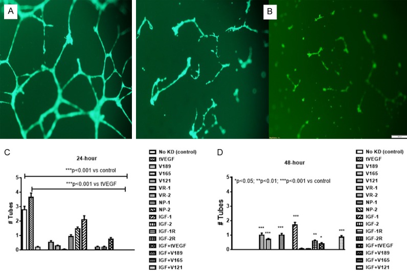
Representative sample of tube formation capacity of human retinal endothelial cells (HRECs) at 24 (A) and 48 (B) hours post transfection. Cells were resuspended in serum-free media and seeded onto 96 well plates. The plates were placed incubated under their growth conditions for 7 days and scored for the presence or absence of spheres at 24 (C) and 48 (D) hours. Data are expressed as mean ± SEM (n=96 samples/group; *P<0.05, **P<0.01, ***P<0.001 vs. control). Images were captured at 20× magnification.