
Figure 2.
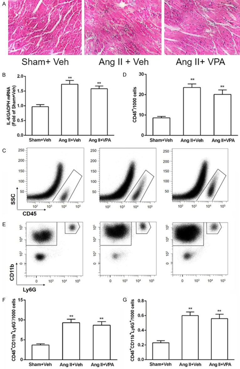
VPA does not reduce Ang II-mediated inflammation in the LV. A: Heart sections of rats treated with Ang II in the absence or presence of VPA were stained by H&E staining, and arrows indicated inflammatory cells. B: IL-6 mRNA expressions was not decreased by VPA treatment. C: Cells falling within the FSC/SSC gate was analyzed for CD45+ positivity against SSC. D: Quantification of CD45+ cells in rat LV, n = 3/group. E: CD45+ cells were then further analyzed for expression of CD11b and Ly6G to identify the monocyte/macrophage and neutrophil populations. F and G: Quantification of indicated populations of monocytes/macrophages (CD45+CD11b+Ly6G-) and neutrophils (CD45+CD11b+Ly6G+) in LV single-cell suspensions, n = 3/group. Data represent mean ± SEM. **P < 0.01 vs Sham + Vel group.