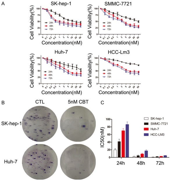
Figure 1.

Effect of cabazitaxel on HCC cell lines in vitro. A. HCC cells were treated with increasing doses of cabazitaxel (0-50 nM) for 12 h, 24 h, 48 h, 72 h. Then, HCC cells proliferation was assessed by the MTT assay. B. SK-hep-1 and Huh-7 cells’ colony formation with or without 5 nM cabazitaxel treatment. C. The IC50s of cabazitaxel after 24 h, 48 h and 72 h treatment were determined for each cell line; Data are shown as mean ± SD. CTL, control; CBT, cabazitaxel.
