
Figure 5.
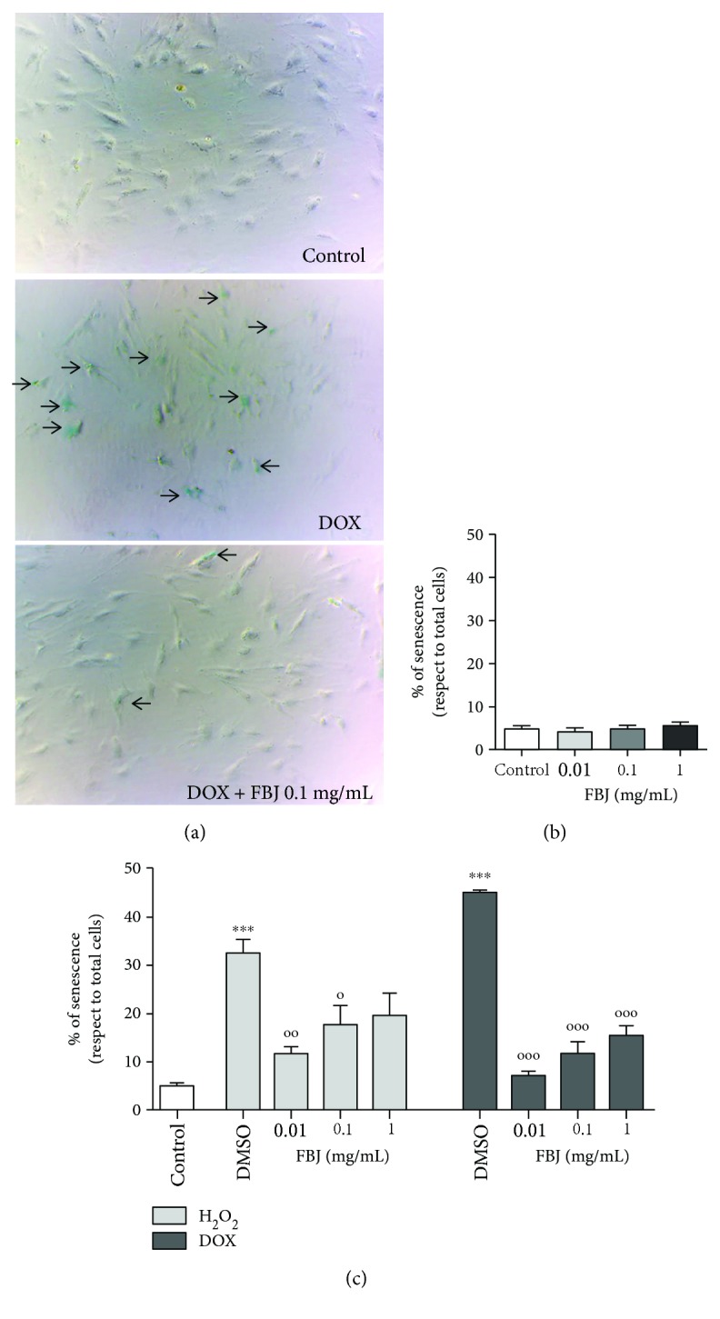
Effects of C. bergamia “fantastico” juice on H9c2 senescence-associated β-galactosidase staining. (a) Representative phase contrast photomicrographs of control cells, DOX-injured cells, and DOX-injured cells treated with FBJ, 0.1 mg/mL. The arrows indicate the blue-stained cells. (b) Percentage of cellular senescence in not-injured cells treated with different concentrations of FBJ. Data are shown as the percentages of β-galactosidase-positive cells with respect to the total cell number of the sample. Each bar represents the mean ± SEM of three replicates from three independent experiments. (c) Percentage of cellular senescence in H2O2- or DOX-injured cells treated with FBJ at different concentrations. Data are shown as the percentages of β-galactosidase-positive cells with respect to the total cell number of the sample. Each bar represents the mean ± SEM of three replicates from three independent experiments. The light gray bars represent the data obtained from the H2O2-induced senescence model; the dark gray bars represent the data obtained from the DOX-induced senescence model. ∗∗∗p < 0.001 versus the control (cells not injured); °°°p < 0.001 versus the H2O2- or DOX-challenged cells with DMSO; °°p < 0.01 versus the H2O2- or DOX-challenged cells with DMSO; °p < 0.05 versus the H2O2- or DOX-challenged cells with DMSO.