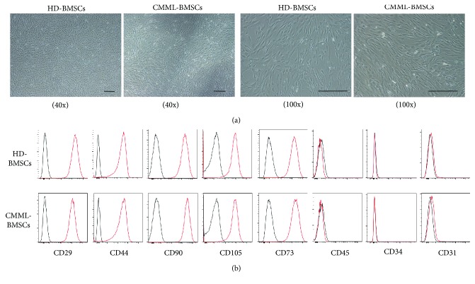
Figure 1.
Characterization of CMML-BMSCs and HD-BMSCs. (a) Typical micrographs depicted the morphology of HD-BMSCs and CMML-BMSCs by light microscopy. Representative images obtained under 40x or 100x magnification are shown. Scale bar indicates 100 μm. (b) Representative figures of cell surface marker analysis in CMML-BMSCs (lower panel) versus HD-BMSCs (upper panel) are shown. The red line represents the BMSCs stained with different antibodies while the black line represents the corresponding negative control stained with isotype-matched nonreactive fluorochrome-conjugated antibodies.

