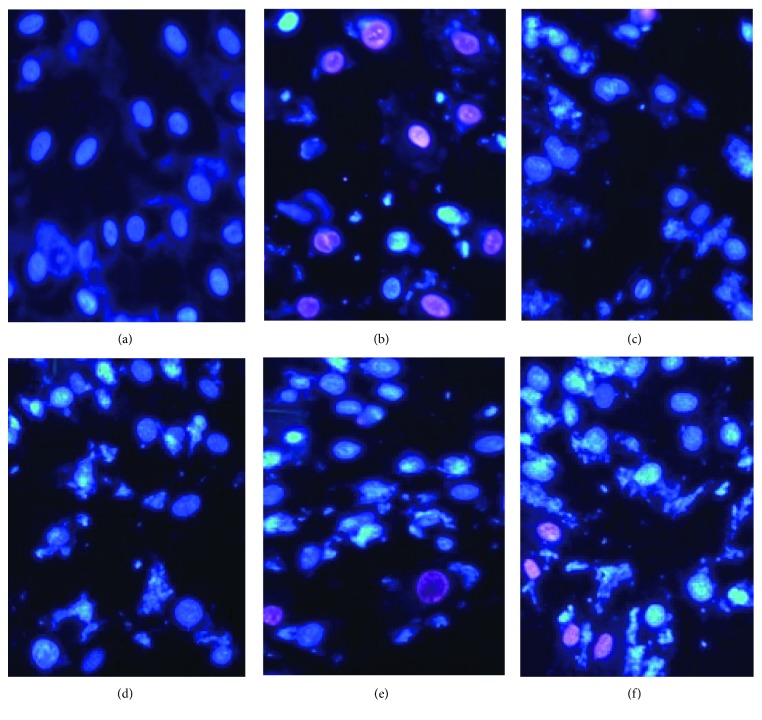
Figure 7.
A double-fluorescent staining kit was used for the detection of the cell cycle and cell necrosis. After Hoechst 33342/PI fluorescent staining, the normal cells, apoptotic cells, and necrotic cells can be distinguished from the flow cytometer (×200). (a) Control group, (b) model group, (c) verapamil 10−11 M group, (d) rosamultin 10−11 M group, (e) rosamultin 10−12 M group, and (f) rosamultin 10−13 M group.

