Figure 5.
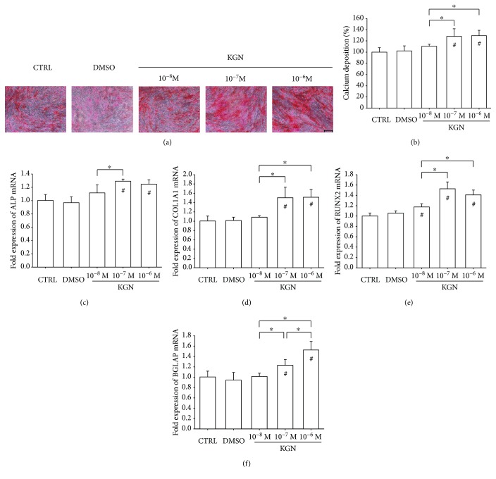
The effect of KGN on osteogenic differentiation of BM-MSCs. Cells were induced toward osteogenic differentiation in the presence of 10−8 M, 10−7 M, and 10−6 M KGN for 14 days. (a) Representative images of mineralized extracellular matrix stained by Alizarin Red S. Scale bar = 200 μm. (b) Quantification of the stained mineral layers demonstrated that KGN increased calcium deposition in differentiated BM-MSCs. The stained mineral layers were treated with perchloric acid, and absorbance was measured at 420 nm. The values were normalized to the level of the CTRL group. (c–f) The mRNA levels of osteoblast-specific marker genes, including ALP (c), COL1A1 (d), RUNX2 (e), and BGLAP (f), were quantified with real-time RT-PCR using GAPDH for normalization. Values are the mean ± SEM of four independent experiments (n = 4) in Alizarin Red S staining and real-time RT-PCR experiments. Statistically significant differences are indicated by ∗ where p < 0.05 between the indicated groups and # where p < 0.05 versus the CTRL group.

