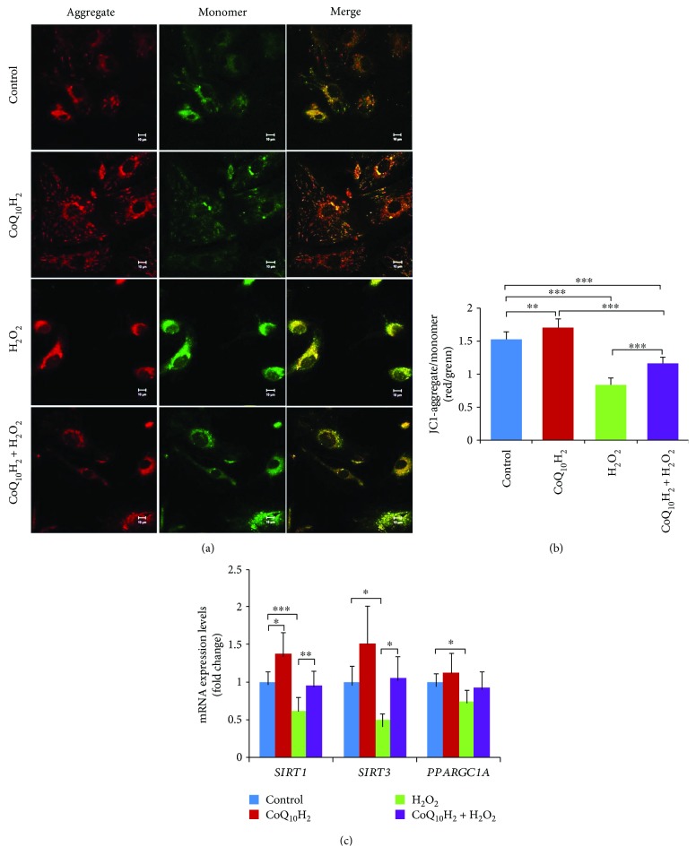
Figure 6.
Preincubation with CoQ10H2 prevents H2O2-induced deterioration of mitochondrial membrane potential in HUVECs. (a) Representative laser scanning microscopy images of aggregated (red) and monomeric (green) JC-1. The two images in each row were captured within the same field and then merged. (b) Mitochondrial depolarization was demonstrated by a change in JC-1 fluorescence from red to green (aggregate/monomer) (n = 6). (c) Real-time RT-PCR analysis of SIRT1, SIRT3, and PGC-1α (PPARGC1A) mRNA expression. Histograms show fold change in mRNA level relative to the control cells (n = 6–9). ∗ P < 0.05, ∗∗ P < 0.01, ∗∗∗ P < 0.001; one-way ANOVA followed by Tukey's test.

