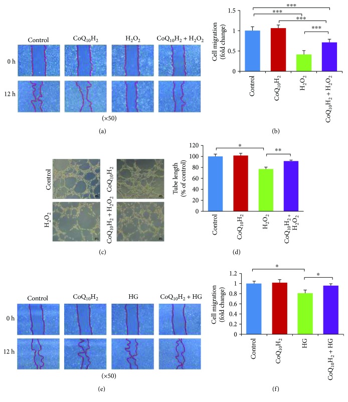
Figure 7.
Preincubation with CoQ10H2 prevented H2O2 or HG-induced reduction in migration and tube formation by HUVECs. (a) Representative images of cell migration analysis evaluated using a wound-healing assay conducted over 12 hours in H2O2-induced reduction of migration. (b) Histograms show fold change in migration activity relative to control cells (n = 9). (c) Representative images from a H2O2-induced tube formation assay after a 6-hour incubation. (d) Histograms show fold change in total cell tube length relative to control cells (n = 6). (e) Representative images of cell migration over 12 hours evaluated using a wound-healing assay in HG-induced reduction of migration. (f) Histogram shows fold change in migration activity relative to control cells (n = 3). ∗ P < 0.05, ∗∗ P < 0.01, and ∗∗∗ P < 0.001; mean ± SD, one-way ANOVA followed by Tukey's test.

