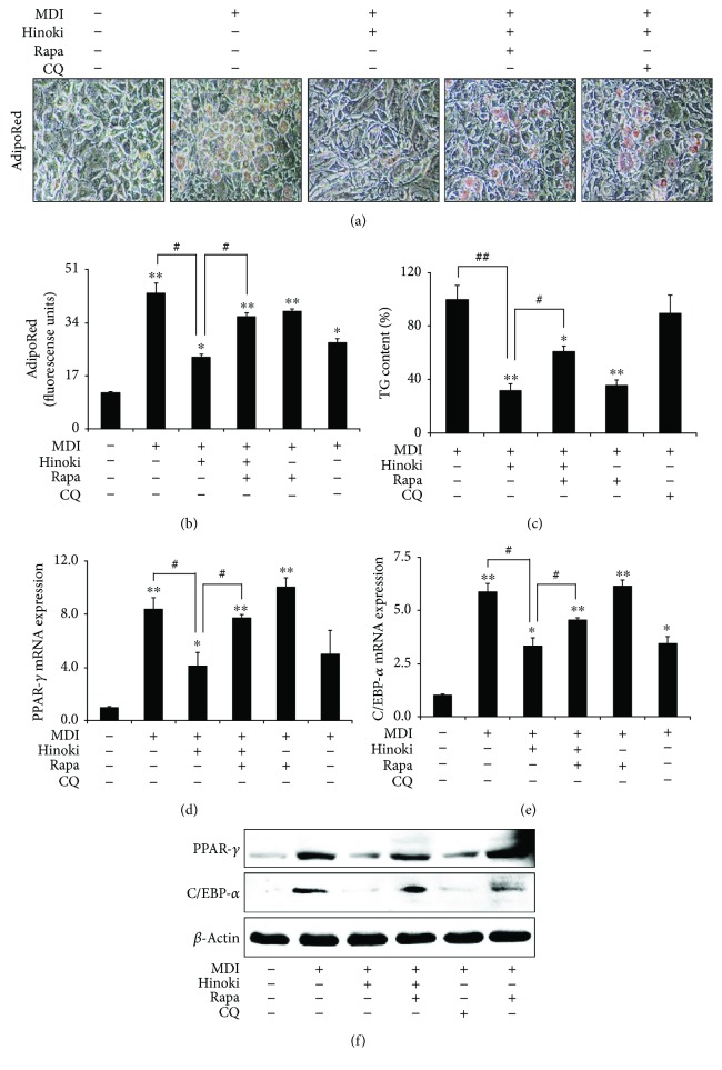
Figure 3.
Induction of autophagy restored inhibited adipocyte differentiation by hinokitiol in mesenchymal stem cells. Mesenchymal stem cells (MSCs) in the presence of CQ or rapamycin for 1 h were incubated with 10 μM of hinokitiol, following treatment with MDI. The AdipoRed assays were performed on day 6 and were photographed with a light microscope (×200). Fluorescence was measured with an excitation wavelength of 485 nm and an emission wavelength of 572 nm (a and b). TG assay (c) was assessed on day 7, and TG contents relative to the control were measured. Total RNA was extracted to quantify the mRNA expression levels of PPAR-γ (d) and C/EBP-α (e). PPAR-γ and C/EBP-α proteins were detected by Western blot analysis (f). β-Actin was used as loading control. Bar graph was generated using mean ± standard error of the mean (SEM) (n = 3). ∗p < 0.05 and ∗∗p < 0.01 for significant differences between the control and treatment groups and #p < 0.05, ##p < 0.01 for significant differences when compared with the LPS treatment group.

