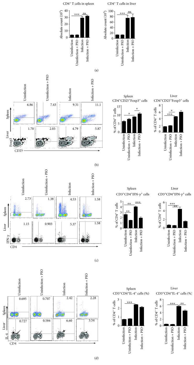
Figure 2.
The expression of regulatory cells and effector Th1/Th2 cells after pioglitazone treatment of S. japonicum infection. (a–d) Single cell suspensions of mouse spleen or liver from pioglitazone-treated mice infected with or without S. japonicum were prepared. (a) The absolute numbers of CD4+ T cells both in the liver and spleen of mice were calculated as the proportion of CD3+CD4+ T cells in the leukocyte gate multiplied by the total cell count. (b) Cells were stained with CD25-APC and CD4-FITC and then intracellularly stained with PE-conjugated antibodies against Foxp3 for FACS analysis of CD4+CD25+Foxp3+ (Treg). (c, d) Cells were stained with CD3-APC and CD4-FITC and then intracellularly stained with PE-conjugated antibodies against IFN-γ or IL-4 for FACS analysis of CD3+CD4+IFN-γ+ (Th1) or CD3+CD4+IL-4+ (Th2) cells, respectively. Data are expressed as the mean ± SD of 12 mice for each group from three independent experiments (ANOVA/LSD), ∗P < 0.05, ∗∗P < 0.01, and ∗∗∗P < 0.001.

