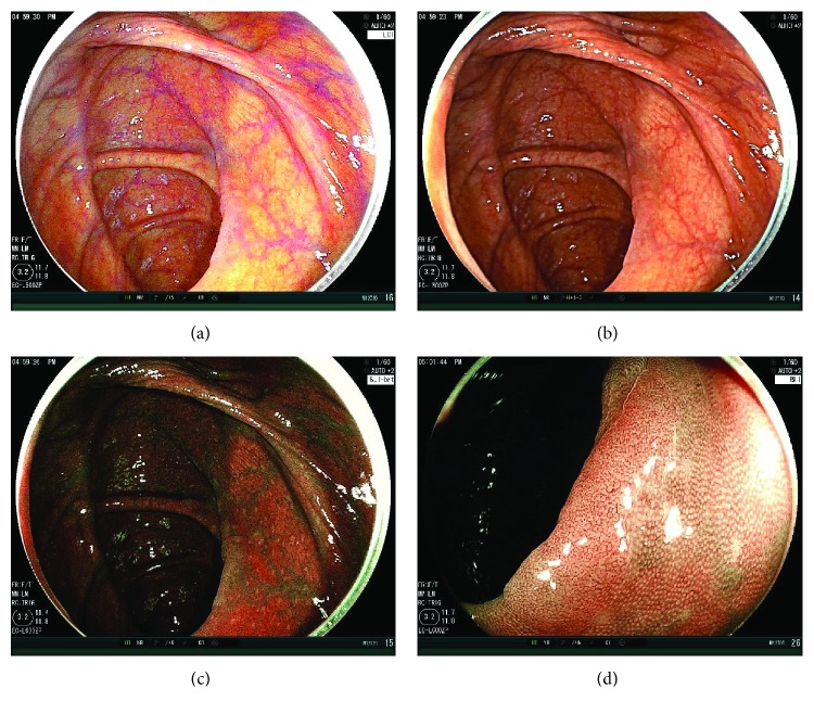
Figure 2.
Case presentation. (a) A nonpolypoid adenomatous polyp 10 mm in size on the ascending colon. The first WLI observation could not detect this tumor. LCI could detect the tumor in a distant view. The endoscopic view was bright, and the tumor was on the fold. It was well-visualized as a little pinkish lesion under LCI compared to the surrounding mucosa. (b) WLI image of this tumor was taken afterwards. The color of it was almost similar to the surrounding tumor. (c) The BLI-bright image of it was also taken. The endoscopic view was a little dark, but the color of the tumor became brownish. (d) BLI magnification showed an adenomatous vessel pattern and surface pattern.

