Figure 3.
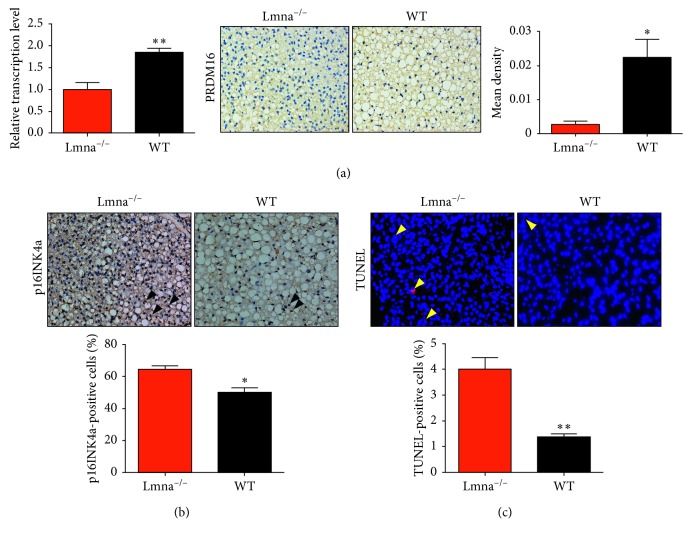
Analysis of the PRDM16 expression levels, and cell senescence and apoptosis rates in the BAT of Lmna−/− and WT mice. (a) Relative transcription levels and immunohistochemical analysis of PRDM16 in Lmna−/− and WT mice at 14 weeks of age. (b, c) Images of p16INK4a immunostaining (black, open arrowheads) and TUNEL (yellow, open arrowheads) in the BAT of Lmna−/− and WT mice and quantification of positive labeling for p16INK4a and TUNEL. ∗ P < 0.05; ∗∗ P < 0.01.

