
Figure 5.
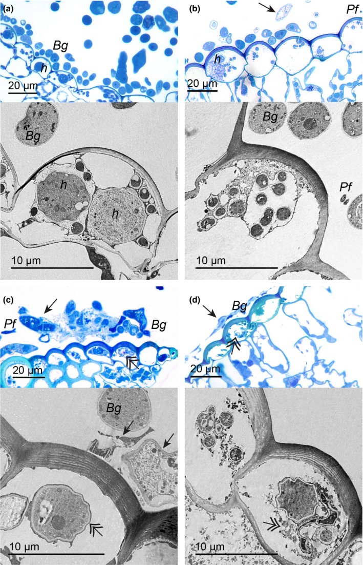
Light and transmission electron microscopy (TEM) of the interaction Pseudozyma flocculosa–Blumeria graminis f.sp. hordei–Hordeum vulgare reveal phenotypic responses supporting transcriptomic results. (a) Light and TEM. Healthy surface hyphae and conidia (Bg), and haustoria (h) of B. graminis are visible in barley cells before inoculation with P. flocculosa (0 hpi). (b) At 12 hpi, presence of P. flocculosa (Pf) as well as B. graminis empty hyphae are observed on the surface (arrows; light), whereas haustoria and digitations are seemingly healthy (TEM). (c) At 24 hpi, surface hyphae and conidia are clearly degraded (arrow; light), in barley cells, the haustoria also show signs of degradation (double arrow; TEM) along with a thicker extra‐haustorial matrix. (d) At 36 hpi, a denser layer of dead hyphae is evident on the leaf surface (arrow; light), and haustorial structures are convoluted when not completely plasmolyzed (double arrow; light and TEM).