
Figure 1.
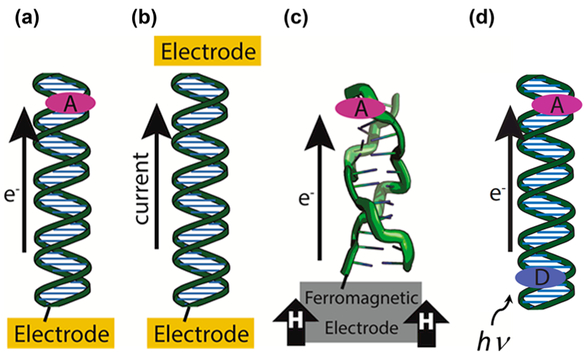
Platforms for the study of DNA-mediated charge transport (DNA CT). (a) DNA is covalently tethered to an electrode surface with an intercalated redox probe. A cyclic increasing or decreasing potential is applied that results in charge being transported through the DNA either to or from the electrode, which can be measured as a change in the current during a potential sweep. (b) DNA is covalently tethered between two electrodes. This type of setup is used to measure the current between the two electrodes in conductive AFM and STM break junction methods. (c) A ferromagnetic electrode influences the yield of charge transport through DNA in different conformations, such as the Z-form shown above. (d) Donor and Acceptor molecules (ovals) are intercalated into a DNA duplex. Transition metal complexes, Ru metallointercalators, Rh metalloinsertors, intercalating organic dyes, and fluorescent base analogs are commonly used as donor and/or acceptor molecules. Photoexcitation initiates charge transport through the DNA bridge and is measured using spectroscopy or other means generally probing the donor or acceptor.