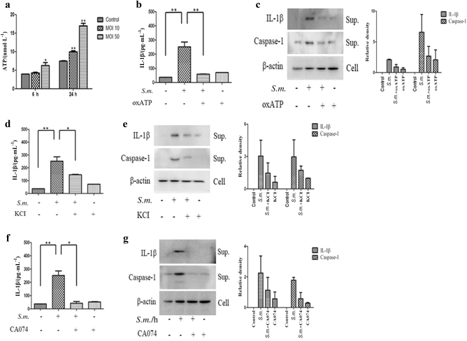
Fig. 4.
ATP release, potassium (K+) efflux, and cathepsin B activity are involved in S. mutans-induced NLRP3 activation. a THP-1 cells were infected with S. mutans (MOI 50) for 24 h. The extracellular ATP concentration was determined using an ATP determination kit. b–g THP-1 cells were pretreated with oxATP (100 μmol · L−1), KCl (500 μmol · L−1), or CA-074Me (5 μmol · L−1) for 30 min before S. mutans infection (MOI 50) for 24 h. Cell culture supernatants were collected and assayed for IL-1β secretion by ELISA (b, d, f). Secreted IL-1β and caspase-1 were detected in the culture supernatant (sup.) by immunoblotting (c, e, g). The results represent those from one of three individual experiments. The data are reported as the means ± standard deviations (n = 3). The relative western blot band densities were normalised to those of β-actin. *P < 0.05, **P < 0.01. S.m. means S. mutans

