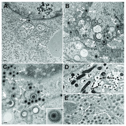
Figure 5. The effect of BFA.
TEM of Vero cells at 9 hpi with R7041(ΔUs3) ( A), at 16 hpi ( B) and at 20 hpi ( C) with wt HSV-1, and at 15 or 17 hpi with wt HSV-1 and BFA exposure ( D and E). ( A) The ER (er) runs from the nucleus (n) towards the cell periphery forming an entity with the PNS that contains a virion (v). ( B) The ER contains virions. One capsid is in the stage of budding (b) into the ER. The ER continues into Golgi (go) membranes at two sites. One Golgi cisterna contains a virion (Vg), one virion has been derived by wrapping (Vw). Close to Golgi stacks, there is probably a virion (V?) of abnormal size. ( C) One capsid buds (b) at the nuclear (n) periphery. The ER is dilated and filled with virions (Ve) and dense material: An ER membrane turns into a Golgi membrane (thick arrow). ( D) After exposure to BFA from 8 to 15 hpi with wt HSV-1, the ER was dilated and contained some virions. ( E) The ER was almost filled with virions after exposure to BFA from 8 to 17 hpi with wt HSV-1. Note that virions in the PNS and ER are covered by a dense coat hiding spikes whereas spikes are clearly apparent on virions in the extracellular space ( C inset). Bars: 200 nm.

