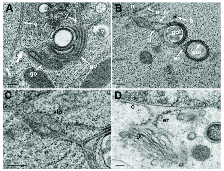
Figure 8. TEM of Vero cells at 12 hpi with R7041(ΔUs3) and of BoHV-1 infected MDBK cells, showing Golgi fields close to the nucleus (n).
( A) Golgi (go) membranes continue (thick arrow) into the ONM (o) as well as towards the cytoplasm indicated by (?) because the destination is unknown. ( B) Golgi membranes continue via ER membranes (er) into the ONM. The ER contains 4 virus-like particles. ( C) Details of panel B. ( D) PNS, ER and Golgi complex form an entity in a BoHV-1 infected MDBK cell ( D: This figure has been reproduced with permission of P. Wild et al., Micron 33, 2002, Elsevier). Bars 200 nm.

