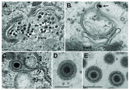
Figure 9. Virions in Golgi cisternae versus budding of capsids.
( A) Golgi cisternae engulfing BoHV-1 virions at 20 hpi. Many of them are tangentially (t) sectioned. ( B) Budding BoHV-1 capsid at a Golgi membrane of the cis-face (arrow). ( C) HSV-1 virion in a Golgi cisterna that connects to the ER (arrow). Note the dense content within the ER and Golgi cisterna indicating little loss of material during processing. ( D) Concentric vacuole derived by wrapping containing a single BoHV-1 virion. The space between viral envelope and vacuolar membrane is always filled in well preserved cells. ( E) Virions in a large vacuole or cisterna exhibiting clearly spikes even in tangentially (t) sectioned virions. Bars: 200 nm.

