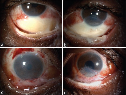
Figure 2.

(a and b) Inferior large area of scleral ischemia with overlying conjunctival defect along with epithelial defect over the inferior one-third of the cornea in both eyes. The tarsal ischemia and lid margin damage are seen. (c and d) Following tenonplasty for scleral ischemiafor both eyes
